


Cocsartrosis- This is the hip joint arthrosis.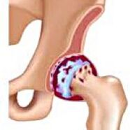 It gradually develops, for years, prone to progression, can be one -one and double -sided.It is accompanied by restrictions on pain and joint movements.In later stages, the atrophy of the muscles of the hip and the abbreviation of the limb can be observed.The diagnosis is determined by clinical symptoms and radiographic results.In the early stages of coxarthrosis, conservative treatment.By destroying the joint, especially for young and middle -aged patients, surgery (endoprostanics) indicates.
General information
The hip joint cokearthrosis (osteoarthosis or deforming arthrosis) is a degenerative-dynist disease.It usually develops at 40 years of age or older.This may be the result of various injuries and joint diseases.Sometimes it occurs for an obvious reason.Coksartrosis is characterized by a gradual progressive course.In the early stages, conservative treatment methods are used.In later stages, the common function can only be restored.
Coxarthosis is one of the most common arthrosis in orthopedics and traumatology.The high frequency of its development is due to the significant load on the hip joint and the widespread prevalence of congenital pathology - joint dysplasia.Women suffer a little more frequently in cocsartrosis than men.
Causes of Coksartrosis
They distinguish between the primary (from unknown reasons) and secondary (as a result of other diseases) arthrosis.
Secondary Coksartrosis may be the result of the following diseases:
- Hip common dysplasia.
- Congenital dislocation of the thigh.
- Pertes diseases.
- The thigh head is aseptic necrosis.
- Infectious injuries and inflammatory processes (such as arthritis of the hip joint).
- Injuries (traumatic dislocations, fracture of hips, pelvic fractures).
Coksartrosis can be one or two -sided.In the case of primary coxarthrosis, simultaneous lesion of the spine (osteochondrosis) and the knee (gonarthrosis) is often observed.
Risk factors
Increasing factors of coxarthrosis are as follows:
- Continuous increased load on the joint.Most often they can be observed in athletes of people with excessive body weight.
- Circulatory disorders, hormonal changes, metabolic disorders.
- Pathology of the spine (kyphosis, skoliosis) or stop (flat foot).
- Elderly and senile age.
- Sitting lifestyle.
Coksartosis itself is not inherited.However, the child can inherit certain characteristics from parents (metabolic disorders, skeleton structural characteristics and weakness of cartilage).Therefore, in the presence of blood relatives with coxarthrosis, the likelihood of the disease has increased slightly.
Patanatomy
The hip joint is made up of two bones: ileum and femur.The head of the thigh is formulated with the acetabulum of the iliac bone and form a specific "wrist strap".During movements, the acetabulum remains motionless and the femur head moves in different directions, ensuring bending, extension, abduction, yield and rotational hips.
During the movements, the joint surfaces of the bones are unobstructed slippage relative to each other, thanks to the smooth, elastic and durable hyaline cartilage that covers the cavity of the rotating cavity and the head of the thigh.In addition, the Hyaline cartilage is a shock -abrasion function and is involved in the redistribution of cargo during movement and walking.
The joint cavity has a small amount of joint fluid that plays the role of lubrication and provides food for the hyaline cartilage.The joint is surrounded by a dense and strong capsule.Above the capsule there are large femoral and gluteal muscles that provide movements in the joint and, along with hyaline cartilage, shock absorbers that protect the joint from injuries with failed movements.
In coxarthrosis, joint fluid becomes thicker and viscous.The surface of the Hyaline cartilage dries, loses smoothness, covered with cracks.Due to the roughness that arises, the cartilage movements are continuously damaged, causing thinning and exacerbating abnormal changes in the joint.As coxarthrosis progresses, the bones are deformed, "adapted" to increased pressure.The joint metabolism deteriorates.In later stages of coxarthrosis, severe atrophy of the painful limb muscles can be observed.
Symptoms of coxarthrosis
The main symptoms of the disease include the joint, inguinal region, thigh and knee pain.In addition, it can be observed on the side of the lesion by cokesartrosis, stiffness of movements and joint stiffness, walking disorders, lameness, and limb abbreviation on the lesion side.A typical feature of coksartrosis is the restriction of kidnapping (for example, the patient is difficult to sit on a chair).The presence of certain signs and their severity depends on the stage of coxarthrosis.The first and the most constant symptom is pain.
-CircleGrade 1 is CoksartrosisPatients complain of periodic pain, which occurs after physical activity (running or long walking).The pain is localized in the joint, less often in the thigh or in the knee.It usually disappears after relaxation.Cape 1, the walking of coxarthrosis is not broken, the movements are completely preserved, there is no muscle atrophy.
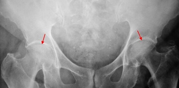
Mild changes are determined in the X scrubbing of a patient with a 1st degree coxartrosis: moderate uneven narrowing of the joint and growth of bones around the outer or inner edge of the acetabulum, in the absence of the femur head and neck.
-CircleCoksartrosis 2 degreesThe pain becomes more intense, often appears at rest, radiating into the comba and groin.After significant physical activity, a patient with coksartrosis begins to soften.The amount of joint movements is reduced: the thigh is abducted and inner rotation.
For X -Ray images, a significant narrowing of the joint gap (more than half of the normal height) is determined for coxarthrosis.The femur head is slightly upward, deformed and increased in size, and the contours become uneven.Bone growth, with this degree of coxarthrosis, appears not only on the outer edge of the internal but also on the outer edge of the acetabulum and goes outside the cartilage.
-CircleCoksartrosis 3 degreesThe pain becomes constant, and the patients' concerns are not only during the day but also at night.Walking is difficult when moving, a patient with coksartrosis is forced to use a reed.The amount of movement of the joint is sharply limited and the muscles of the buttocks, hips and lower legs were atrophy.The weakness of the thigh removal muscles is the cause of the pelvic difference in the anterior plane and shortens the limb on the sore side.To compensate for the abbreviation, the patient with cokertrosis occurs in the sore direction when walking.As a result, the gravity center is shifted and the strain of the painful joint is suddenly increased.
X -rays at 3 degrees Coxartrosis are sharp reducing the joint gap, outstanding expansion of the thigh head and multiple bone growth.
Diagnosis
Diagnosis of coxarthrosis is based on the clinical symptoms and data of further tests, which are most important to radiography.In many cases, X rails allow not only the degree of coxarthrosis but also the cause of its occurrence.Thus, for example, the growth of the cervical-diaphragm angle, the flattening of the scenes and the acetabulum indicate dysplasia, and changes in the shape of the proximal part of the femur indicate that coksartrosis is a consequence of pertes disease or youth epiphyolic.X -rays of patients with coxarthrosis can also be detected to indicate injuries.
Such as other methods of instrumental diagnosis of coxarthrosis, CT and MRI can be used.Computer tomography allows you to examine pathological changes in detail and magnetic resonance imaging allows you to evaluate the disorders by soft tissues.
Differential diagnosis
First and foremost, coxarthrosis should be distinguished from gonarthrosis (osteoarthosis of the knee joint) and osteochondrosis of the spine.The atrophy of the muscles, which occurs in the 2 and 3 stages of Coxarthosis, can cause pain in the knee joint, which are often brighter than pain in the area of injury.Therefore, with the patient's knee pain, clinical (control, palpation, determination of the amount of movement) is to study the hip joint and, if it is assumed that coxarthrosis is directed to the patient to X -ray.
Radicular syndrome (compression of nerve roots) may imitate pain with coxarthrosis for some other diseases of the spine.Unlike cocsartrosis, the pain occurs when the roots are pressed suddenly, after a failed movement, sharp turn, lifting weight, etc., is localized in the buttocks and spreads to the back of the thigh.Positive symptoms of tension are observed - severe pain when the patient tries to compensate for a straightened limb, lying on his back.At the same time, the patient is free to take his leg, while in patients with coksartrosis, the abduction is limited.Keep in mind that osteochondrosis and coksartrosis can be observed at the same time, so thorough examination of the patient is always required.
In addition, cokesartrosis can be distinguished by trochanteritis (boot busite) - aseptic inflammation in the area of gluteal muscles.Unlike coxarthrosis, the disease develops quickly within 1-2 weeks, usually after injury or significant physical activity.The intensity of the pain is higher than in the case of coksartrosis.Movement restrictions and shortening of the limb are not observed.
In some cases, the atypical course of the disease or reactive arthritis can be observed with symptoms similar to coxarthrosis.Unlike coxarthrosis, these diseases, the peak of pain fall at night.The pain syndrome is very intense and can be reduced while walking.Morning stiffness, which is immediately awakened and gradually disappears within a few hours.
Treatment of coxarthrosis
The treatment of pathology deals with traumatologist orthopedics.The choice of treatment methods depends on the symptoms and stage of the disease.Coxarthrosis 1 and 2 sections are performed by conservative therapy.During the aggravation of coxarthrosis, the injection blocks, non -steroidal anti -inflammatory drugs (pyroxians, indomethacin, diclofenac, ibuprofen, etc.) are used.Keep in mind that the drugs of this group are not recommended for a long time as they can have a negative impact on the internal organs and suppress the capability of hyaline cartilage.
To restore the cocsartrosis of damaged cartilages, the funds of a group of chondroprotectors (chondroitin sulfate, cartilage extract, etc.) are used.Vasodilation drugs (zinnarisin, nicotinic acid, pentoxifillin, xantinol nicotinate) are prescribed to improve blood circulation and eliminate small blood vessels.According to indications, muscle relaxants (muscle relaxation drugs) are used.
Patients with stubborn pain syndrome can be prescribed intraarticular injections using hormonal drugs (hydrocortisone, triamcinolone, metrum).Treatment with steroids should be done with caution.In addition, local products are used in addition to coxarthrosis - heating ointments with no pronounced therapeutic effect, but in some cases relieve muscle cramps and reduce pain due to their "disturbing" effect.In addition, physiotherapy procedures (lighting, ultrasonic therapy, laser treatment, UHF, inductance, magnetic therapy), massage, manual therapy and therapeutic gymnastics are prescribed.
The diet of coksartrosis does not have an independent therapeutic effect and is only used as a means of weight loss.Reducing body weight allows you to reduce the load on the hip joints and, as a result, facilitates the course of coksartrosis.To reduce the load on the joint, your doctor may recommend that you go with reeds or crutches, depending on the degree of coxarthrosis.
In later stages (with degrees 3 coxarthrosis), the only effective treatment method is surgery - replacing the destroyed joint with endoprothesis.Depending on the nature of the lesion, the only -pole (replacing the head of the thigh only) or two -poles (replacing the head of the thigh and rotating cavity) can be used.
Endoprostication is performed for coxarthrosis as planned, after full examination, after general anesthesia.In the postoperative period, antibiotic therapy is performed.The seams are removed for 10-12 days and the patient is prescribed for outpatient treatment.Rehabilitation measures are necessarily retained after endoprostics.
In 95% of cases, surgical intervention to replace the joint with coxarthrosis ensures complete restoration of the limb function.Patients can work, actively move and even exercise.The average service life of the prosthesis, with each recommendation, is 15-20 years.Thereafter, a second operation is needed to replace worn endoprothesis.